HIDA Scan: What to Know About the Diagnostic Tool
Medical imaging has revolutionized how healthcare providers diagnose and treat various conditions, particularly those affecting the digestive system. Among these diagnostic tools, the HIDA scan stands as a specialized imaging test that, while less commonly known than x-rays or CT scans, provides valuable insights into specific digestive functions. This nuclear medicine procedure has become increasingly important in helping doctors identify and diagnose certain gastrointestinal conditions that other imaging methods might miss.
What Is a HIDA Scan?
A HIDA scan, which stands for hepatobiliary iminodiacetic acid scan, is a specialized imaging test that helps healthcare providers examine the function of certain digestive organs. Also known as hepatobiliary scintigraphy or cholescintigraphy, this procedure belongs to a category of tests called nuclear medicine. Nuclear medicine procedures use small amounts of radioactive materials called tracers to create detailed images of internal organs and their function, making them different from standard x-rays that only show organ structure.
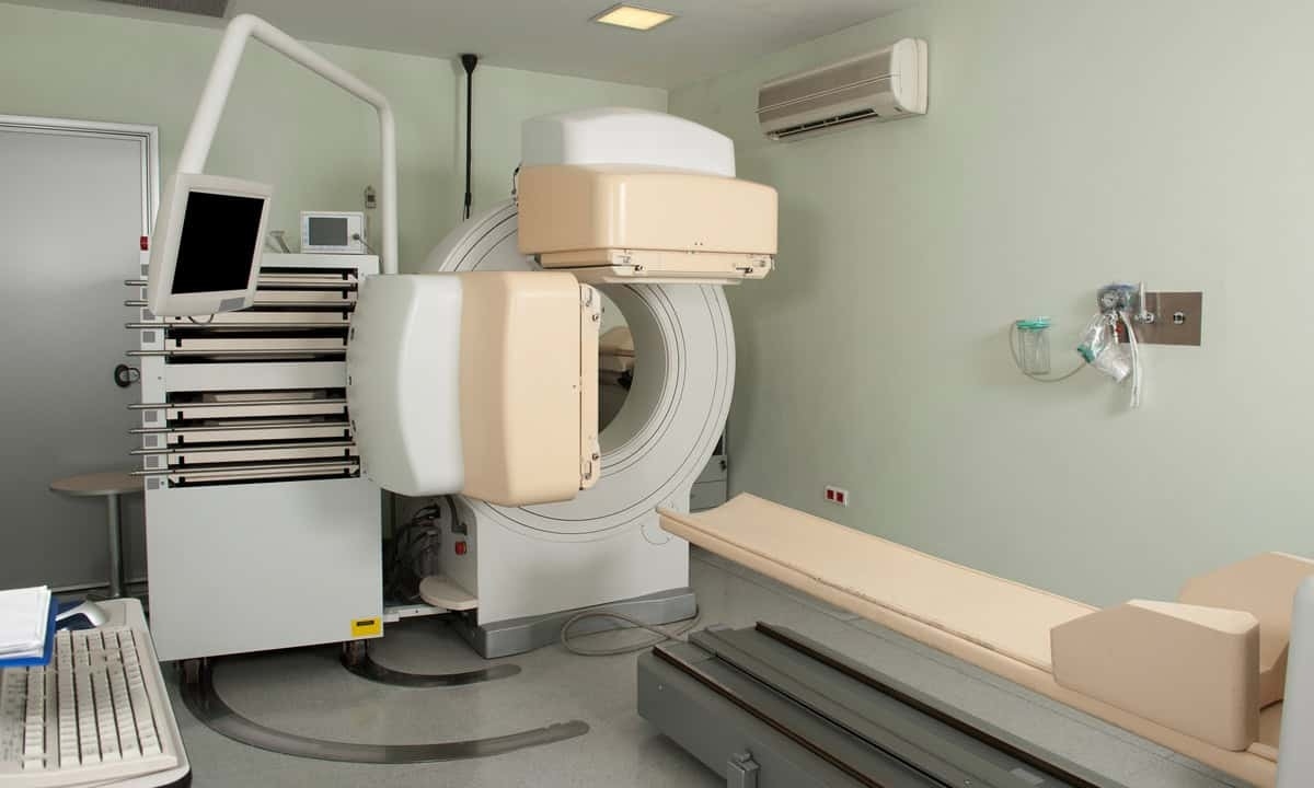
During a HIDA scan, a technologist injects a radioactive tracer into a vein in the patient's arm. This tracer is designed to travel through the liver, bile ducts, gallbladder, and small intestine—collectively known as the biliary system. A special camera called a gamma camera tracks the tracer’s movement through these organs, allowing doctors to observe how well this system is functioning. The tracer’s journey through the body provides valuable information about bile flow and organ function that other imaging tests cannot capture.
The name “HIDA” might sound intimidating, but it simply refers to the type of radioactive tracer used in the scan. The amount of radiation exposure during a HIDA scan is very low and similar to what you might receive from common imaging tests. The procedure is performed in a radiology or nuclear medicine department by specially trained healthcare professionals who carefully monitor the entire process.1
Reasons to Perform a HIDA Scan
Healthcare providers often recommend HIDA scans when they suspect problems with the biliary system, particularly issues affecting the gallbladder or bile ducts. These scans are especially useful for identifying conditions that affect how bile flows through the digestive system. Bile, a substance produced by the liver, plays a crucial role in digestion and needs to flow freely through various organs to function properly. Common reasons for performing a HIDA scan include:
- Acute cholecystitis: A HIDA scan can identify sudden inflammation of the gallbladder, often caused by gallstones blocking bile ducts.
- Chronic cholecystitis: The scan helps diagnose ongoing gallbladder inflammation that may develop over time.
- Bile duct blockages: HIDA scans can detect obstructions preventing normal bile flow through the biliary system.
- Gallbladder dysfunction: By measuring the gallbladder ejection fraction using cholecystokinin (CCK), the scan can show how effectively the gallbladder contracts and releases bile.
- Post-surgical evaluation: The scan can identify potential bile leaks following surgery or check the placement of biliary stents.
- Pre-transplant assessment: Doctors may use HIDA scans to evaluate patients before or after liver transplant procedures.
- Unexplained symptoms: The scan can help investigate severe abdominal pain on the right side or unexplained jaundice when other tests are inconclusive.
What to Expect During a HIDA Scan
A HIDA scan takes place in a hospital’s radiology or nuclear medicine department and typically requires several hours to complete. The process often begins with patients changing into a hospital gown to keep personal clothing from interfering with the imaging. A nuclear medicine technologist then places an intravenous (IV) line in the arm to administer the radioactive tracer, which will travel through the biliary system during the test.
During the scan itself, patients lie still on an examination table while a gamma camera moves slowly around them, tracking the tracer’s journey through the body. The camera takes a series of images as the tracer travels from the liver through the bile ducts and into the gallbladder and small intestine. Some patients may receive an injection of cholecystokinin (CCK) partway through the test to measure gallbladder function, which can cause temporary feelings of nausea or discomfort similar to being very hungry.
Most patients find the HIDA scan to be a relatively comfortable procedure, though remaining still for extended periods can be challenging. While allergic reactions to the tracer are rare, healthcare providers carefully monitor patients throughout the test. Patients may need to avoid eating for a period before the scan, and certain medications or supplements might need to be temporarily discontinued. The nuclear medicine team will provide specific preparation instructions based on individual circumstances.2
Interpreting the Results of a HIDA Scan
After the scan is complete, a radiologist analyzes the series of images showing how the radioactive tracer moved through the biliary system. The pattern and timing of the tracer’s flow can reveal various conditions. For example, if the tracer doesn’t enter the gallbladder, this might indicate inflammation or blockage. Similarly, if the tracer moves too slowly or appears in unexpected areas, it could signal problems with bile flow or potential leaks.
HIDA scan results help healthcare providers identify specific issues like gallbladder inflammation, bile duct blockages, or gallbladder dysfunction. During part of the test, doctors measure how well the gallbladder contracts and empties itself. If the gallbladder doesn’t empty properly, this may indicate it isn’t working as it should, even when other imaging tests show the gallbladder looks normal. This information helps doctors determine the best course of treatment for patients experiencing gallbladder-related symptoms.
Following the scan, patients can immediately resume normal activities. The small amount of radioactive tracer naturally leaves the body within a few days through normal bodily functions. While patients might need to temporarily limit close contact with pregnant women and young children as a precaution, the low radiation exposure during a HIDA scan is comparable to that of many common imaging tests. Healthcare providers will discuss any specific aftercare instructions based on individual circumstances and test findings.
Contact Cary Gastro for More Info
While HIDA scans are just one of many diagnostic tools available, they provide valuable information about gallbladder and bile duct function that other imaging tests can’t capture. If you’re experiencing symptoms like severe abdominal pain, particularly on your right side, the experienced gastroenterologists at Cary Gastro can help determine if a HIDA scan might be appropriate. Our team works closely with imaging specialists to ensure accurate diagnosis and effective treatment of biliary system disorders. Contact us today to request an appointment and discuss your symptoms with our healthcare team.
1https://www.ncbi.nlm.nih.gov/books/NBK539781/
2https://www.ucsfhealth.org/medical-tests/gallbladder-radionuclide-scan