Endoscopic Ultrasound: Advanced Imaging for Digestive Health
Medical imaging technology continues to evolve, providing doctors with increasingly detailed views of internal organs and tissues. Endoscopic ultrasound (EUS) represents a significant advancement in diagnostic capabilities, particularly for examining the digestive tract and nearby organs. This specialized procedure combines traditional endoscopy with high-frequency sound waves, allowing gastroenterologists to examine areas that other imaging methods, like CT or MRI, might not capture as clearly.
What is an Endoscopic Ultrasound?
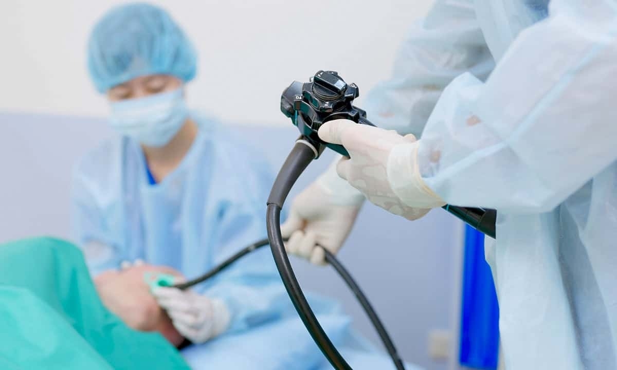
Endoscopic ultrasound combines two powerful imaging technologies into a single procedure. A gastroenterologist uses a specialized flexible tube (endoscope) with an ultrasound device built into its tip. This instrument allows for both direct visualization of the digestive tract through traditional endoscopy and detailed ultrasound imaging of nearby structures using high-frequency sound waves.
The endoscope can be inserted either through the mouth or rectum, depending on which areas need examination. For an upper gastrointestinal tract endoscopic ultrasound, the scope passes through the mouth, esophagus, and stomach into the first part of the small intestine (duodenum). Lower endoscopic ultrasound involves passing the scope through the rectum to examine the colon. As it moves through the digestive tract, the ultrasound device produces detailed images of the gastrointestinal tract wall and surrounding tissues.
Unlike traditional ultrasound performed from outside the body, endoscopic ultrasound can position the imaging device very close to the organs being examined. This proximity allows for extremely detailed images that help identify small abnormalities in tissue structure. The procedure can also guide fine needle aspiration (FNA) when tissue samples are needed for further testing, making it especially useful for targeted biopsies.
EUS is particularly valuable for examining organs like the pancreas, which traditional imaging methods might struggle to visualize clearly. The high-frequency sound waves used in EUS create detailed images of pancreatic tissue, helping doctors identify and evaluate various conditions, from inflammation to tumors. Similarly, endoscopic ultrasound provides excellent visualization of the bile duct system, gallbladder, and lymph nodes surrounding the gastrointestinal tract, often revealing abnormalities that other methods miss.1
Reasons for Performing an Endoscopic Ultrasound
An endoscopic ultrasound helps healthcare providers evaluate various conditions affecting the digestive system and nearby organs. The detailed images it provides can identify problems that other imaging tests might miss, making it particularly valuable for both diagnosis and treatment planning. In some cases, doctors might recommend an endoscopic ultrasound after other imaging studies, such as tomography scans or cholangiopancreatography (ERCP), have shown unclear or concerning results. Common reasons for performing an endoscopic ultrasound include:
- Pancreatic conditions: Evaluating pancreatic tumors, cysts, or chronic pancreatitis, often providing more detailed images than other imaging methods. EUS can detect very small abnormalities in the pancreas and help determine whether they require treatment.
- Digestive tract abnormalities: Examining suspicious areas in the esophagus, stomach, or other parts of the gastrointestinal tract. The procedure helps determine the depth of tumors and whether they have spread to nearby lymph nodes.
- Cancer staging: Determining how far certain cancers may have spread and whether they have affected nearby lymph nodes. This information proves crucial for planning appropriate treatment strategies.
- Bile duct problems: Investigating blockages or other issues affecting the bile duct system. EUS can help identify the cause of jaundice or unexplained abdominal pain related to the biliary system.
- Tissue sampling: Guiding EUS-guided fine needle aspiration (FNA) to obtain tissue samples from suspicious areas for further testing. This targeted approach helps ensure accurate sampling of concerning areas.
- Treatment planning: Helping determine the best approach for treating various gastrointestinal conditions. The detailed images can guide decisions about surgery, medication, or other interventions, and can be particularly helpful in planning the placement of stents if needed to relieve blockages.
What to Expect During an Endoscopic Ultrasound
Before an endoscopic ultrasound, patients receive specific preparation instructions from their healthcare provider. For upper EUS, patients typically fast for 6-8 hours before the procedure to ensure clear imaging. Lower EUS might require similar preparation to a colonoscopy, including dietary restrictions and taking solutions to clean the colon. The procedure takes place in a hospital or outpatient endoscopy center, where the medical team can monitor patients closely throughout the process.
During the procedure, patients receive sedation to ensure comfort. The type and level of sedation depend on various factors, including the specific examination needed and individual patient characteristics. The gastroenterologist then gently guides the endoscope through either the mouth or rectum, depending on which areas need examination. The ultrasound device at the tip of the scope produces sound waves that create detailed images of internal organs and structures. If needed, the doctor can perform a fine needle aspiration to collect tissue samples for further testing.
The entire endoscopic ultrasound procedure typically takes between 30-90 minutes, depending on the areas being examined and whether tissue samples are collected. Throughout the process, the medical team carefully monitors the patient’s vital signs and comfort level. The combination of sedation and the skill of experienced gastroenterologists helps ensure the procedure is well-tolerated.2
Post-Procedure Care
Following an endoscopic ultrasound, patients spend some time in a recovery area while the sedation wears off. The recovery period usually lasts 30-60 minutes, during which time nurses monitor vital signs and ensure patients are becoming alert. Most people can return home the same day, though they’ll need someone else to drive them due to the lingering effects of sedation. Recovery is typically quick, with most patients returning to normal activities the next day.
Common side effects after the procedure are usually mild and temporary. These might include a slight sore throat if the endoscope was inserted through the mouth, or minor bloating and gas. While rare, more serious complications can include bleeding, infection, or perforation, particularly when tissue samples are taken. The healthcare provider carefully explains potential warning signs and provides clear instructions about when to seek immediate medical attention.
Results from the endoscopic ultrasound imaging are often available quickly, though tissue sampling results may take several days. The healthcare team schedules appropriate follow-up care based on the findings and may recommend additional testing or procedures if necessary. Most patients find the procedure much easier than anticipated and appreciate the valuable information it provides for their care.
Contact Cary Gastro for More Information
An endoscopic ultrasound provides valuable insights for diagnosing and treating various digestive conditions. If you’ve been recommended for this procedure, the experienced gastroenterologists at Cary Gastro can help. Our team specializes in performing EUS procedures with the latest technology and techniques, ensuring you receive the highest quality care in a comfortable setting. Contact us today to request an appointment and learn more about how endoscopic ultrasound might benefit your digestive health.
1https://pmc.ncbi.nlm.nih.gov/articles/PMC4952290/
2https://pancan.org/facing-pancreatic-cancer/diagnosis/endoscopic-ultrasound-eus/